Understanding the Colorless Nature of Viruses
Written on
Chapter 1: The Color of Viruses
Viruses are devoid of color, not even gray. They are indifferent to our advanced human perception.

Viruses are not red
Viruses are not blue
Magnify them as you will
They remain invisible to you!
Viruses are minuscule entities, and calling them "creatures" is somewhat misleading, as they do not qualify as living organisms. Unlike bacteria, which are single-celled life forms, viruses depend entirely on a host's cellular machinery for their existence and replication.
Let’s delve into their size and color. The dimensions of viral structures typically range from 100 to 400 nanometers. For instance, coronaviruses measure approximately 120 to 160 nanometers.
Bacteria, by comparison, are larger, measuring around 1000 nanometers or 1 micrometer. So, what does their small size imply? While they are indeed microscopic, they are even tinier than what an ordinary optical microscope can detect, regardless of its magnification capabilities.
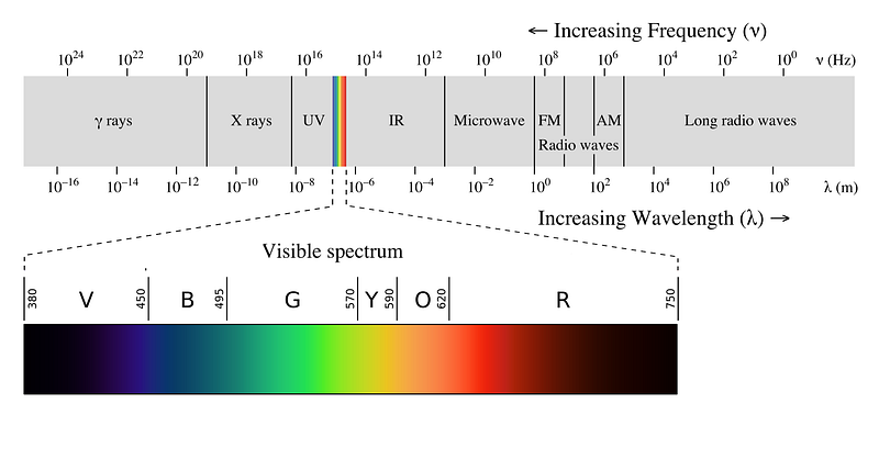
At these scales, we approach the lower threshold of visible light reflection. Visible light is an electromagnetic wave characterized by specific wavelengths. The shortest visible wavelength is roughly 400 nanometers, which our eyes perceive as violet-blue. Conversely, wavelengths around 700 nanometers appear red to us.

All colors visible to us are electromagnetic waves with wavelengths between 400 and 700 nanometers (±50 nanometers). The perception of color is a biological construct, shaped by human evolution in response to the sunlight's spectrum.
For more insights on how nature might appear on other planets, refer to this article. Now, let's continue.
From the perspective of the universe, there is no inherent distinction between visible light and other electromagnetic waveforms. The primary difference lies in the energy carried by the wave; shorter wavelengths correspond to higher frequencies and greater energy. For example, blue light possesses more energy than red light, while X-rays carry even higher energy than visible light.
Given that a virus measures about 120 nanometers, it falls below the shortest visible wavelength. Consequently, it cannot reflect any colors we recognize. The electromagnetic wave simply passes around it, rendering it invisible, no matter how advanced the microscope.
You might bring a chicken to a microscope, but you can’t compel it to look.
Interestingly, some creatures, like birds and chickens, have vision adapted for detecting shorter wavelengths, such as ultraviolet light. If you were to place a chicken under a powerful microscope, it might be able to perceive the virus, while we would not, due to its ability to detect electromagnetic waves outside our visible spectrum.
However, for a virus as diminutive as 120 nanometers, it would require an ultraviolet vision capability. This wavelength is already close to the X-ray range. I doubt we have chickens with X-ray vision, but who knows—perhaps a superhero could see it.

So how can we humans, with our limited vision, observe these tiny viruses? Instead of utilizing photons for visual detection, we can employ electrons through an electron microscope (EM). Electrons, while different from photons, share wave-particle characteristics. The wavelength associated with an electron is approximately a thousand times shorter than that of visible light, roughly 1 nanometer.
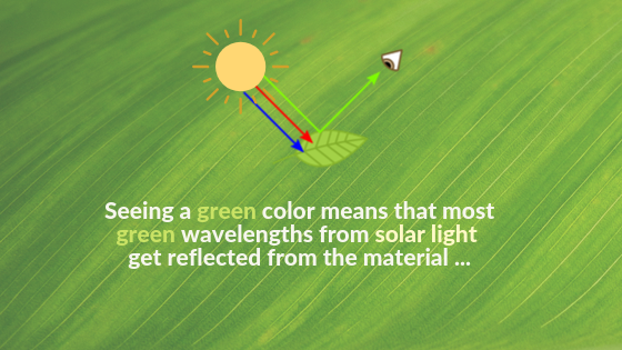
When electrons are directed at a virus under specific conditions, some will bounce back, allowing us to visualize the virus. This process is analogous to how light interacts with objects; when light strikes a surface, some is absorbed while some is reflected, defining the color we perceive.

Similarly, electron microscopes function by using electrons instead of photons. Since electrons are charged and would dissipate in air, the sample must be placed in a vacuum. Instead of optical lenses bending light, electromagnetic coils manipulate the trajectory of electrons in an EM.
For more detailed information on electron microscopy techniques, check out this comprehensive guide: Electron Microscopes explained.


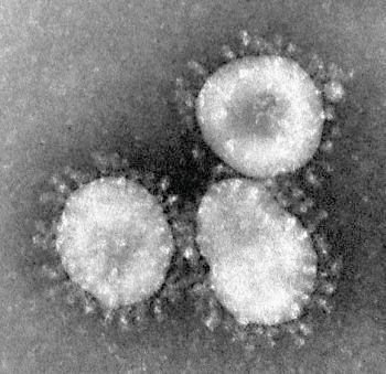
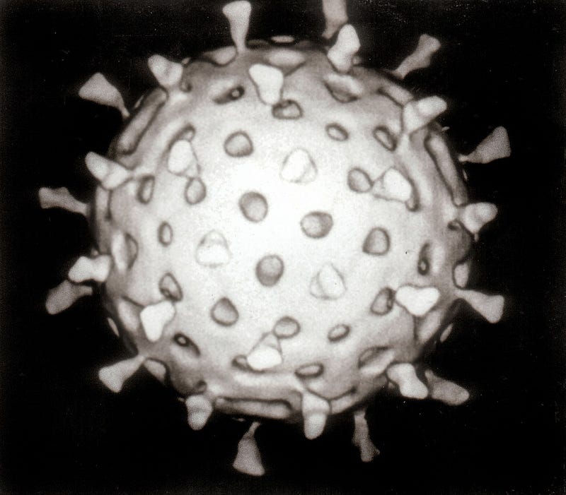
Left: A coronavirus captured by an electron microscope. The name stems from the crown-like appearance of the virus. Image source: Wikimedia. Right: A rotavirus visualized in an electron microscope, enhanced with computer graphics. Image source: Wikimedia.
Viruses are not “gray” either.
The gray-scale images produced by electron microscopy do not imply that viruses possess a gray color. In fact, they are fundamentally colorless, reflecting no visible light, including wavelengths perceived as gray. This gray appearance in EM images often results from visual enhancement or computer-assisted rendering to clarify their structures.
Next question to ponder: Do viruses have any smell or taste?
Chapter 2: Insights into Virus Visualization
The first video, titled "How to Fix Discord Stuck on a Gray or Black Screen [Still Works on 2024]," guides viewers through resolving common display issues in Discord.
The second video, "This MAGIC Vitamin ACTUALLY IS Repigmenting My Gray Hair (truly shocked)," shares the surprising results of a vitamin that claims to restore natural hair color.